|
Overview:
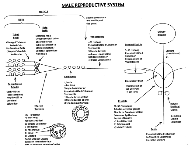
The goal of this lab is to examine the histology of the principal parts of the male reproductive tract. Use the text referenced above and the diagram below as a guide. Understand the functional correlates of the architecture you observe, and be able to distinguish each of the components. Pay attention to the disposition of the epithelium, note the special modifications of tubules and glands, and observe the amount and layering of connective tissue and smooth muscle.
Slide Descriptions
I. TESTIS
270 testis testis H&E Webscope Imagescope [Orientation]
270ex testis H&E Webscope Imagescope [Orientation]
275 Testis H&E Webscope Imagescope [Orientation]
UCSF 363 testis Webscope Imagescope
These slides (except for the UCSF slide) include both testis and epididymis (you will study epididymis later in this laboratory session): the testis is the larger of the two structures on the slide. The capsule of the testis is composed of dense connective tissue and is called the tunica albuginea. Within the testis you will see numerous profiles of seminiferous tubules, with interstitial tissue between them. In the interstitial tissue between the seminiferous tubules are clusters of Leydig cells, which secrete the male steroid hormone, testosterone. These are the most prominent cells in the interstitial tissue (best seen in #270 [example], or #275 [example]). Also visible in the interstitial tissue are blood vessels and smaller cells characteristic of loose connective tissue.
Now take a closer look at the the seminiferous tubules in #270 [example] [Orientation] or #270 [example] [Orientation] (in some of the seminiferous tubules the epithelium may be pulled away somewhat from the basement membrane, leaving a white space, which is an artifact). Each seminiferous tubule is surrounded by a boundary layer or tunica propria, composed of flattened cells, several cells thick. Most of the cells that lie against the basement membrane and have round nuclei are spermatogonia. Don’t worry about distinguishing between type A and type B spermatogonia. Now look for considerably larger nuclei midway up in the epithelium that are also round and are filled with a tangle of dense chromosomes. These are the nuclei of relatively mature primary spermatocytes, which are in the extended prophase of the first meiotic division. These are numerous but unfortunately not always well preserved (the nuclei may be somewhat swollen or distorted).
All of the smaller cells in the upper half of the epithelium (toward the lumen of the tubule) are spermatids in various stages of differentiation into mature sperm. The spermatids initially have round nuclei, but these gradually become smaller, denser and assume the shape of sperm heads. The smallest black structures you see are the heads of mature spermatids about to be released into the lumen. Their tails are difficult to make out.
Now look for Sertoli cell nuclei, which are large, relatively pale and irregular in shape, and contain a prominent nucleolus (sometimes out of the plane of section). Their nuclei commonly occur just above (toward the lumen from) the spermatogonia. Secondary spermatocytes are rarely seen, since almost immediately after they arise they undergo the second meiotic division to become spermatids. If you are anxious to see secondary spermatocytes, find division figures and look around them for nuclei that are intermediate in size between those of primary spermatocytes and spermatids. They are hard to find, so don’t spend much time looking. How can you differentiate between Sertoli cells, spermatogonia and primary spermatocytes?
II. MEDIASTINUM and EPIDIDYMIS
270 testis testis H&E Webscope Imagescope [Orientation]
270ex testis H&E Webscope Imagescope [Orientation]
275 Testis H&E Webscope Imagescope [Orientation]
Returning to the slides of the seminiferous tubules, study the passageway by which the sperm pass from the seminiferous tubules through the rete testis, efferent ducts and epididymis to reach the tail of the epididymis, where they are stored in preparation for an ejaculation. Scan along the tunica albuginea of the testis, looking for a region where it thickens and is permeated by a network of flattened channels in the dense connective tissue. This portion of the testis is known as the mediastinum; the network of channels is the rete testis and is probably best seen in slide #270 [example] or #275 [example]. Observe that the rete testis is lined with a cuboidal (or sometimes low-columnar) epithelium and you may see occasional microvilli. (Purely as an aside, these cells also have a single or “primary” cilium. Obviously, these cilia don’t contribute much in the way of helping to propel sperm through the channel. Instead, they may function as chemoreceptors allowing the lining cells to monitor and modify the luminal contents).
The sperm leave the testis by means of these channels to reach the efferent ducts. However, for now, shift your attention outside the testis to the part of the epididymis that is included in this slide. You will see numerous sections through the long and convoluted duct that makes up the epididymis apparent in #275 [example] and #270 [example] [CAVEAT] . The epithelium of the duct is pseudostratified columnar. The tall epithelial cells have long microvilli on their apical surface (sometimes called “stereocilia”, although they are not cilia at all). You may be able to make out a thin layer of smooth muscle around the tubules, which presumably acts by peristalsis to move the sperm along.
Now scan over the section looking for tubules of the efferent ducts, which connects the rete testis to the epididymal duct. The efferent duct tubules are often (but not always) smaller than epididymal tubules, and their epithelium varies considerably in height, giving the tubule lumen a characteristic irregular or “star” shape. The epithelium is generally simple columnar, and consists of two cell types, taller cells have with cilia and shorter cells without cilia. The efferent duct is the only portion of the male tract displaying true, motile, cilia. These tubules may also have a thin layer of smooth muscle around them. Efferent ducts are best demonstrated in #275 [example], although #270 [example] shows a small region of efferent ducts amongst the epididymis.
III. SPERMATIC CORD
284 spermatic cord H&E Webscope Imagescope
This is a cross section through a spermatic cord, such as you will see in the gross anatomy laboratory. On one side of the section you will see a cross section of the ductus (or vas) deferens [example], which has a circular lumen, lined by a pseudostratified epithelium and surrounded by a very thick wall of smooth muscle. Elsewhere you will see sections through two or more branches of the testicular artery, surrounded by connective tissue that contains numerous veins of the pampiniform plexus [example] (the veins are rather flattened and contain dark red blood). What is the function of the pampiniform plexus?
The testicular artery has a typical internal elastic membrane and prominent circular smooth muscle coat (comprising the tunica media). The veins of the pampiniform plexus lack the internal elastic membrane, and their walls are somewhat less muscular. The blood in the vessels may have black particles in it, an artifact of the fixation procedure. The association of the veins of the pampiniform plexus with the testicular artery constitutes a countercurrent exchange system to cool the blood somewhat on its way to the testis. In the connective tissue you will find nerves and, around the periphery of the section, some cross-sectioned bundles of smooth (dartos) [example] and skeletal (cremaster) muscle [example].
IV. SEMINAL VESICLE
075 seminal vesicle H&E Webscope Imagescope
279 seminal vesicle H&E Webscope Imagescope
These are cross sections through a seminal vesicle, which you may remember from gross anatomy is a rather sacculated and contorted tube (slide 75 is also the slide we used to study parasympathetic ganglia, which are readily observable in the wall of this organ). Study the section with your light microscope, noting the moderately-abundant smooth muscle in the wall. The epithelium of this gland lies on the surface of interconnecting mucosal folds that extend into the lumen from the muscular wall. The sparse connective tissue within the folds constitutes the lamina propria of this mucosa. The epithelium, which may be either simple columnar or pseudostratified columnar, produces a secretion (including fructose, ascorbic acid and other components) which is expelled from the gland by contraction of the muscular wall during ejaculation, constituting about 50-80% of the semen.
V. PROSTATE GLAND
281 prostate H&E Webscope Imagescope [Orientation]
281lex prostate Masson Webscope Imagescope
282 prostate senile H&E Webscope Imagescope
The prostate in an adult man is about the size and shape of a chestnut and contains 15-30 tubuloalveolar glands that empty separately into the prostatic urethra. The most important structures to see on the slide are the prostatic glands that are present over most of the section. The glands are embedded in a connective tissue that includes abundant smooth muscle, best appreciated in the trichome-stained section [example] (recall that collagen fibers will stain blue-green whereas smooth muscle is pink with deep red nuclei), which produces the pulsations of the prostate that expel the content of the glands during ejaculation. The epithelium of the glands is simple columnar (there may be a few basal cells), and the cells differ greatly in height to give the epithelium a folded appearance. The epithelial cells secrete various components of the semen (including citric acid and acid phosphatase). In older individuals, there may be concretions (amyloid bodies) in the lumen of the glands, which are better seen in #282 [example] –the concretions are of little functional consequence, but they can be a helpful for the purposes of identifying the tissue. The prostatic urethra can also be seen in slide 281 [example] (it has a stratified epithelium several cells thick and numerous mucosal glands). Also present on slide 281 is the utricle flanked by paired ejaculatory ducts [example].
With these landmarks, one can identify the various zones of the prostate that are clinically relevant in their propensity to become hyperplastic and/or cancerous:
- The peripheral zone [example] contains the main glands located posterio-laterally in the outer parenchyma of the gland and is the site most susceptible to inflammation (prostatitis) and malignant neoplasia (prostatic carcinoma).
- The central zone [example] is the region surrounding the utricle and ejaculatory ducts. Interestingly, this zone is relatively resistant to inflammation and hyperplasia.
- The transitional and periurethral zones [example] surrounds the prostatic urethra and contains submucosal and mucosal glands that undergo non-cancerous proliferation, leading to benign prostatic hypertrophy (BPH), a condition that effects almost all males to some extent by age 80. Typically, the submucosal glands [example] undergo hypertrophy first followed by the mucosal glands as the condition progresses.
- The anterior fibromuscular stroma [example] is located anteriorly and contains no glandular tissue so it typically does not become hyperplastic.
VI. PENIS
286 penis H&E cross Webscope Imagescope [Orientation]
This is a cross section through the shaft of the penis. Hold the slide up to the light and identify the two pale corpora cavernosa [example] (occupying most of the central part of the section; the partition between the two may be incomplete), surrounded closely by a thick layer of dense connective tissue (pink), the tunica albuginea. Also observe the corpus spongiosum [example], situated ventromedially and containing the slit-like penile urethra. Study the erectile tissue of the corpora cavernosa with your microscope, noting the connective tissue trabeculae and smooth muscle strands, as well as small blood vessels and nerves. The arterioles within the trabeculae are called helicine arteries(oles) [example] because in the flaccid penis they are rather twisted (like a helix). Between the trabeculae are the venous sinuses, lined with endothelium. In the flaccid penis, the smooth muscle in the trabeculae and arterioles is under tonic contraction, limiting the entry of blood into the sinuses. Erection involves a relaxation of the smooth muscle of the arterioles (so more blood enters) and of the trabeculae (permitting the sinuses to expand freely), as well as a closing down of venous outflow due to compression of the veins that drain the erectile tissue (the veins draining the tissue are pushed up against the rigid connective tissue of the tunica albuginea when the erectile tissue swells). The organ expands by hydraulic pressure, and the shape of the erect penis is determined by the tunica albuginea. A resumed contraction of the smooth muscle in the helicine arterioles leads to detumescence. In each corpus cavernosum, note one or two arteries, the deep arteries [example], that supply the arterioles. If you look on the upper surface of the tunica albuginea, you will see the abundant nerve bundles that supply this organ.
|