The goal of this lab is to examine and describe the structural organization of the heart and the major classes of blood vessels.
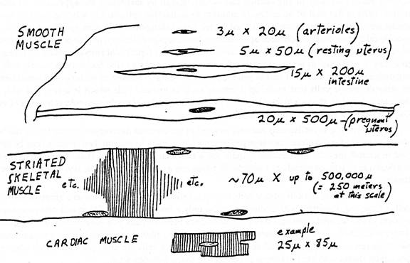
Below is a schematic drawing showing the key structural features and relative sizes of skeletal, smooth, and cardiac muscle as you would observe them with high power.
Slide Descriptions
I. Heart
A. Cardiac Muscle: Webslide 0026_B: Cardiac Muscle, monkey, H&E [DigitalScope]
Cardiac muscle fibers can be seen in both cross and longitudinal sections. Measure fiber diameters and note blood vessels filled with RBCs between the fibers. Fine details of the myocardiocytes can be seen, especially myofibrils, cross striations, intercalated disks, and mitochondria next to the nuclei.
Note also the simple squamous epithelium covering both free surfaces of the heart in this section. How can you tell whether each surface is endocardium or epicardium? Based on a low-power view, can you tell whether this section was taken from an atrium or a ventricle?
Slide 98HE is stained with H&E and Slide 98-N is a similar section stained with Aldehyde Fuchsin-Masson. You should look at both stains. Locate the atrioventricular sulcus that contains a branch of the coronary arterial system (a muscular artery that exhibits moderate intimal thickening) embedded in the epicardial fat. Look at the connective tissue present between the ventricle and atrium. This is part of the cardiac skeleton into which cardiac muscle inserts. A leaflet of an A-V valve takes origin from the cardiac skeleton. Look at the atrial and ventricular endocardium, consisting of an endothelial lining and the underlying connective tissue (the endothelium is often stripped away during processing, but there are some areas where it has been preserved). With low power, locate the Purkinje fibers present immediately beneath the ventricular endocardium in the H&E[example] and trichrome-stained[example] sections (note the appearance of these fibers in cross and longitudinal orientations). These conducting fibers are larger and paler staining than the cardiac muscle fibers. Note the meshwork arrangement of the cardiac muscle fibers in the myocardium. These slides also offer excellent views of capillaries within the myocardium [example].
The lumen of the ventricle is covered by a simple squamous epithelium and an underlying layer of connective tissue. Deep to this connective tissue is a thick layer containing Purkinje fibers. Measure the diameter of the Purkinje fibers and their nuclei. Are intercalated discs present in these fibers? Note the paucity of fibrils within these cells. What pale staining material fills the central portion of these fibers?
The interventricular septal connective tissue is present, and, in most sections, a distinct unit of specialized cardiac muscle, the A-V bundle (of His), traverses the septal connective tissue (a thin group of muscle fibers surrounded by dense c.t.). The A-V bundle is easiest to see in slide 99HM, [example], although you should also be able to recognize it the H&E-stained section[example] as well. In these slides, the bundle fibers are cut in cross section and they are similar in size and staining to that of normal cardiac muscle fibers, although in some of your sections the fibers may more closely resemble Purkinje fibers (which is what they are). On one side of the section, a leaflet of the aortic valve[example] is present. On the other side, portions of an A-V valve[example] are present, as are bits and pieces of collagenous chordae tendinae. In slide 99H&E, there is a piece of chorda tendinae actually attached to the valve [example] , whereas in slide 99M, the pieces are unattached and out in the ventricular lumen (the attachment site is out of the plane of section [example] .
II. Blood Vessels
Type of Vessel
Outer Diameter (Approx.)
Intima features
Media features
Adventitia features
Roles in Circulatory System
Elastic arteries
> 10 mm
Endothelium; connective tissue with smooth muscle
Many elastic lamellae alternating with smooth muscle
Connective tissue, thinner than media, with vasa vasorum
Conduct blood from heart and with elastic recoil help move blood forward under steady pressure
Muscular arteries
10-1 mm
Endothelium; connective tissue with smooth muscle, internal elastic lamina prominent
Many smooth muscle layers, with much less elastic material
Connective tissue, thinner than media; vasa vasorum maybe present
Distribute blood to all organs and maintain steady blood pressure and flow with vasodilation and constriction
Small arteries
1-0.1 mm
Endothelium; connective tissue less smooth muscle
3-10 layers of smooth muscle
Connective tissue, thinner than media; no vasa vasorum
Distribute blood to arterioles, adjusting flow with vasodilation and constriction
Arterioles
100-10 µm
Endothelium; no connective tissue or smooth muscle
1-3 layers of smooth muscle
Very thin connective tissue layer
Resist and control blood flow to capillaries; major determinant of systemic blood pressure
Capillaries
10-4 µm
Endothelium only
A few pericytes only
None
Exchange metabolites by diffusion to and from cells
Venules
10-100 µm
Endothelium; valves sometimes present
Pericytes and scattered smooth muscle cells
None
Drain capillary beds; site of leukocyte exit from vasculature
Small veins
0.1-1 mm
Endothelium; valves sometimes present; connective tissue with scattered smooth muscle fibers
Thin, 2-3 loose layers of smooth muscle cells
Connective tissue, thicker than media
Collect blood from venules
Medium veins
1-10 mm
Endothelium; connective tissue, with valves
3-5 more distinct layers of smooth muscle
Thicker than media; longitudinal smooth muscle may be present
Thickest layer, with bundled longitudinal smooth muscle
Return blood to heart
A. Muscular (medium) artery and veins
Webslide 0174_B: Muscular Artery & Vein [DigitalScope]
On the right side of WebSlide 174 is a muscular artery and its companion vein. Focusing on the artery first, note the prominent internal and external elastic laminae, which are stained pink in this slide. The tunica media contains several layers of circumferential smooth muscle mixed with collagen. The tunica adventitia which is slightly thinner than the tunica media grades into surrounding fatty connective tissue. The companion vein is just below and the the left of the artery. Note the thickness of the wall compared to the overall diameter of the lumen.
Slides 42 and 95M are mesentery spreads that contain numerous excellent examples small muscular arteries and their companion veins. Study the arteries first in slide #42[example] , and/or #95 (trichrome)[example] . Note the thin intima, the distinct internal elastic lamina (IEL) and the media composed of circularly oriented smooth muscle cells. The media also contains some elastin and abundant “reticular” collagen, as well as specific proteoglycans. There is sometimes a condensation of elastic fibers in the outer portion of the media that may be an “external elastic lamina”, which varies from artery to artery. The adventitia is the dense, irregular connective tissue surrounding the media that varies in thickness. Remember that in blood vessels the components of the media are arranged circularly, while those of the adventitia are oriented longitudinally. In vessels where the media is too thick to be supported by simple diffusion, vasa vasorum (e.g. shown here in slide 95M) may also be observed in the adventitia.
The structure of the companion veins in slide #42[example] and/or #95 (trichrome) [example] is less regular and may be difficult to understand at first, but still consists of the same basic layers as arteries with a tunica intima, media, and an adventitia, although the media is usually much less muscular and less organized compared to the companion artery. Conversely, the adventitia is usually thicker in veins and may often have some bundles of longitudinal smooth muscle[example] as well as vasa vasorum [example] . In some sections, you may be able to see a thin internal elastic lamina beneath the venular endothelium (particularly in slide #95M).
B. Arterioles and venules: Webslide 0098_B: Urinary Bladder, monkey, H&E [DigitalScope]
Use this section to locate many small and medium sized blood vessels. Several good examples of arteries, veins, and capillaries can be observed within the dense connective tissue in the upper left region of the slide. In particular, examine carefully the series of arteries and veins about 50 to 100 mm in diameter that are located about one-third of the way across the slide from the left edge of the section. Identify as specifically as possible each of these vessels based on the amount of smooth muscle in relation to the vessel diameter. Also note the valves in one of the veins.
C. Capillaries
Refer to sections of the HEART above (e.g. Slide 98HE [DigitalScope]) to see examples of capillaries, which are abundant within the myocardium of the heart. Typical capillaries are just small enough to allow single red blood cells (~7um in diameter) to pass.
E. Aorta: Webslide 0022_B: Aorta, monkey, H&E [DigitalScope]
This is an example of an elastic artery. Note:
Tunica intima with endothelial nuclei, longitudinal subendothelial fibers of reticulin and smooth muscle.
Tunica media with multiple wavy light pink elastic membranes (up to 70 such laminae may be counted in a human aorta) and interleaving circular smooth muscle fibers.
Tunica adventitia with some loose fatty areolar connective tissue and inner dense irregular collagen containing vasa vasorum.
Think about possible effects of normal blood pressure distension on endothelial cell plumpness and intimal corrugation. You might expect more penetration by vasa vasorum in thicker vessels of larger animals.
E. Vena Cava / Large Vein: Webslide 0023A_B: vena cava, monkey, plastic, c.s., H&E [DigitalScope]
This is a portion of the inferior vena cava cut in cross section. The original circumference of 10-15 mm is only partly sampled in your slide.
Note the following:
Variable intima with some longitudinal fibers, including smooth muscle [example].
Disorganized media consisting of 3-5 layers of circular smooth muscle) [example].
Thick adventitia with plentiful longitudinal smooth muscle bundles like islands in a sea of collagen [example], with vasa vasorum[example] and surrounding connective tissue --note that the connective tissue of the adventitia here has frayed apart somewhat, so a lot of the empty spaces seen here are due to this artificial separation.
The following additional Webslides may be used to review this material:
A 55-year-old man complaining of chest pain was admitted to the hospital. His clinical evaluation quickly led to the diagnosis of myocardial infarction, primarily involving the left ventricle. The patient appeared restless, anxious, and markedly dyspneic. He appeared pale and his extremities were cool to the touch. Bradycardia was present and the patient was hypotensive. Shortly after admission, the patient developed pulmonary edema and liver failure and unfortunately died a week later. Slide 32 is from the patient's liver.
Once you've thought of the answer to the question above, click here to see what the lungs look like. The field of view that opens is an alveolus which should normally be filled with just air, but there is clearly more than just air here. The large cells are macrophages stuffed with glossy brown pigment.
What is this pigment and where did it come from?
In terms of hemodynamics, what is happening in the lungs?
These sections of arteries came from a 62-year-old hypertensive diabetic who had generalized lesions of this sort.
Focus on the section on the right side of the slide [example] since it's easier to make out what's left of the original lumen of the vessel, which is lined by a simple squamous endothelium [example].
About 2/3 of the original lumen has been taken up an athersclerotic plaque, within which you can see "foam cells" [example] that have ingested cholesterol.
The cholesterol accumulation is so great that it also collects extracellularly as large, elliptical "crystals" (also called cholesterol clefts) [example] --of course, since they are lipid, they are extracted with tissue processing, so they appear as empty spaces.
Within the plaque, you may also see amorphous, basophilic debris [example], which are examples of dystrophic calcification.
What are the possible fates of this sort of lesion?
This 63-year-old patient had a “heart attack” and died shortly thereafter. The immediate cause of his death was an irreversible cardiac arrhythmia.
First, we'll focus on an area of healthy myocardium [example]. Be sure you can identify it as cardiac muscle.
Next, we'll move into the area of the infarct[example], which is a bit darker staining. The increased eosinophilia is due to protein coagulation within the dead cardiomyocytes. The areas of basophilia are streams of neutrophils coming in as part of the acute inflammatory process. Zoom in on an area within the infarct [example] and note that the general structure of the cardiomyocytes is preserved, but the nuclei in the necrotic cells are faded or completely absent (karyolysis) and the cytoplasm is markedly more eosinophilic compared to normal cardiomyocytes [example] and the extracellular space around the necrotic cells is filled with inflammatory cells.
What type of inflammatory cells are these?
About how much time had elapsed between the time of the infarction and the time that this sample was obtained?
Can you predict in general histologic terms what would have happened to this tissue over time had this patient survived? (Slide 49 [DigitalScope] illustrates some of this.) What are some of the complications that the patient might have had to deal with?
A 19-year-old student presented to the campus health service complaining of transient knee and hip joint pains of one week’s duration. He had become febrile 2 days prior to seeking medical attention. Physical examination revealed swollen knee joints and small painless subcutaneous swellings located in the scalp and over the elbows. One week later, a heart murmur thought to be due to mitral regurgitation was detected. Congestive heart failure soon developed and the patient succumbed despite aggressive medical treatment. Slide 50 is prepared from the autopsy material.
What epicardial abnormality is present [hint] and how might this have been clinically manifested on physical examination of the living patient?
Valvular endocardial fibrin deposits (excrescences) are present in the slide [example]. What would these deposits have looked like grossly? If the patient had survived, what would be the clinical significance of these endocardial fibrin deposits?