Epithelial Tissue |
||
| Text: | ||
Overview: The primary goal of this lab is to learn how to identify the various classes of epithelial tissues found in the body and the apical specializations of specific epithelial types. A second, critical goal is to correlate function with structure. Thus identification of these epithelial types and specializations will provide direct information on the function of the tissue and organ.
I. Epithelial Tissue Webslide 0098_G: Urinary Bladder, monkey, H&E This slide contains a section of the wall lining the urinary bladder. When you look at the webslide, the lumen of the bladder is at the bottom and the body cavity is at the top of the field. At low power, contrast the appearance of the natural "free edges" at the bottom and top of the section where epithelium lines the bladder lumen and the body cavity, respectively, from the sharp "cut edges" at the left and right of the section where the tissue was cut during specimen preparation. First focus on the top surface of the tissue, and note where a simple squamous epithelium called a mesothelium forms part of the peritoneal covering of the bladder. This cellular layer is quite thin (less than 5 mm). Note the bulging nuclei and attenuated cytoplasm that forms a continuous sheet that is sharply differentiated from the underlying connective tissue and muscle. Some portion of the specimen has lost its mesothelium, so scan along the top edge of the webslide. Another place to look to find simple squamous epithelium is in the lining of blood vessels, where it is called an endothelium. Many of the blood vessels in this specimen show the endothelial lining quite well. Next examine the lower portion of the slide which is the tissue that lines the lumen of the bladder. Notice the characteristics of the transitional epithelium that is found only in the urinary tract: (1) there are several layers of cells, (2) most of the cells in the outer layer touching the lumen are rounded or polygonal and contain spherical nuclei, (3) in this outer layer of cells there is usually a considerable amount of cytoplasm between the nuclei and the apical plasma membrane. Contrast this with stratified squamous epithelium where the outer layer of living cells is ALWAYS flattened and ALWAYS has thin, flattened nuclei.
Webslide 0038_J: Submandibular gland, monkey,T.B-A.F, 1.5 µm sec. Most of this section is composed of darkly staining acini (balls or clusters of cells) that comprise the parenchyma (dominant cell type) of this organ. Our focus here is on the relatively small, pale staining ducts that can be found at low power within the connective tissue separating the acini. Look for rings of pale-staining cells surrounding a small lumen. These ducts are lined by simple cuboidal epithelium. Note the size and shape of these cells and their central, round nucleus.
Webslide 0032_G: Ileum, monkey, H&E The absorptive surface lining the lumen (bottom of webslide) is thrown into large folds (villi) which increase the absorptive surface area. Scan the slide to observe the orientation of the simple columnar intestinal epithelial cells that line the lumen as they are cut in different planes. Two types of epithelial cells are present in this columnar epithelium – absorptive cells with striated (brush) borders and secretory goblet cells. Within the epithelium, you may also notice some small, dark nuclei scattered about –these are actually lymphocytes, which we will learn more about when we study connective tissue in the next lab session. Identify the following features of the columnar epithelium and understand their fine structure and function:
UMiss_198_40x: Fallopian tube (oviduct isthmus), human H&E. The lumen of the oviduct (near the center of the slide) is thrown into many folds that are all lined by a simple columnar epithelium. Scan the epithelial surface to find regions where the epithelium is cut in a section perpendicular to the basal lamina. Two distinct types of columnar cells are present in this epithelium, ciliated cells and non-ciliated secretory cells. Measure the height of the cilia and compare to the height of the striated border composed of microvilli in the previous Webslide 32. Can you resolve individual cilia at highest power?
UMich_126alt_40x: Esophagus, human, H&E Starting with a low magnification objective, scan the entire bottom surface of the slide and note the non-keratinized stratified squamous epithelium lining the esophagus which contains several layers of cell. The inner layers (away from the lumen) contain cuboidal or polygonal cells, whereas the outer layers (lining the lumen) contain flattened cells with flattened nuclei.
Webslide 0065_G: Skin, Foot, human, H&E The epidermis of the skin, found at the bottom of the slide, is an example of keratinized stratified squamous epithelium. There is a change in cell shape from cuboidal at the base of the epithelium to flattened at the surface. The outermost layers (i.e. at the surface of the skin) consist of keratinized plates containing no nuclei.
Webslide 0026_R: Trachea, human, H&E The epithelium of the trachea is along the bottom of the slide. Note the different levels of the nuclei in this pseudostratified columnar ciliated epithelium. The elongated nuclei of the columnar ciliated cells are farther from the basal lamina than the rounded nuclei of the short basal cells. Goblet cells can also be observed with their aggregated mucinogen granules near the surface of the epithelium. Observe and measure the dimensions of the cilia. Cilia are involved in transport of material along the epithelial surface. What do they transport in the trachea?
II. Pathology Correlate Webslide 222418: Esophagus, human, biopsy, H&E The main purpose of showing this slide is to demonstrate a phenomenon known as metaplasia, whereby an epithelium transforms from one type to another in response to repeated injury The specimen on this slide is from a patient that had a 10-year history of gastro-esophageal reflux disease (GERD), a condition in which stomach contents "reflux" or come back up into the esophagus. This is perceived as an intense burning sensation localized to the chest, hence the common term, "heartburn." In response, to these repeated injuries, the esophagus undergos changes commonly known as Barrett esophagus (named for the surgeon, Norman Barrett, who described the condition in 1950). Approximately 10% of patients with chronic GERD will exhibit these changes. Look along the upper surface of the slide [example] and you'll see a fairly typical stratified squamous non-keratinized epithelium similar to the esophageal lining you observed in Slide 126alt above. (A subtle difference that you may note is that blood vessels under the epithelium are more engorged due to the irritation --see if you can find the endothelium in these vessels...) Now, track leftward and you will see the tissue suddenly transtitions to a very different type of epithelium [example]. See if you can classify what type of epithelium it is. In this patient, the lining of the esophagus has responded to repeated exposure to stomach acid by transforming into an intestinal type of epithelium that is better able to resist the acid environment (mucus and bicarbonate secreted by the goblet cells is protective). Since the transformation is into an intestinal type of epithelium, this is specifically intestinal metaplasia. Since the transformation of the epithelium entails "reprogramming" the stem cells of this tissue (i.e. to become intestinal-type cells rather than squamous esophageal-type cells), these lesions are not surprisingly at higher risk of developing into cancer. The overall risk of these lesions becoming cancerous is about 1% per year; its incidence increases with duration of symptoms and increasing patient age. Although many esophageal cancers are associated with a history of Barrett esophagus, it should be noted that MOST individuals with Barrett esophagus do NOT go on to develop esophageal cancer.
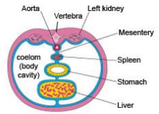
Extra Slides (for additional practice) Here are some additional slides to get some more practice looking at epithelial tissue: Slide UMich #30: Abdominal mesentery, human, H&E
As shown in this figure above, the abdominal mesentery is a membranous sheet that envelops all of the internal organs and suspends them within the abdominal cavity. Just as the organs are covered by a mesothelium, so too is the mesentery. Look all along the lower edge of the specimen and observe the simple squamous epithelium (mesothelium). Within the mesentery are numerous blood and lymphatic vessels which are lined on their inner surfaces with simple squamous epithelium called endothelium.
Slide UMich #009-N1: Kidney, human, H&E This is a section of the kidney and thus features structures involved in urine production, the most prominent of which are the many tubules, lined by simple cuboidal epithelium. In some areas (particularly the upper left hand corner) the tubules are cut more in longitudinal section whereas in other areas the tubules are more in cross section. You may also note variations in the degree of staining (some of the tubules are lined with pale cells while others are lined by much more eosinophilic cells). The more eosinophilic cells are much more involved in active transport and therefore have many more microvilli and mitochondria (which stain strongly with eosin) compared to the paler cells.
Slide UMich #153: Esophagus, human, H&E The esophagus is lined by a non-keratinized stratified squamous epithelium. This type of epithelium covers some internal surfaces that are kept moist by mucus or other fluids, and the lubrication provided by mucus helps to protect against abrasion. Thus, these epithelia do not need to keratinize to resist desiccation and mechanical wear. Study this type of epithelium and note how the cell morphology changes from the roughly cuboidal in the basal layer to squamous in the apical surface of the epithelium (in this case, the apical surface is the bottom-most edge of the tissue). Recall that stratified epithelia are named based on the morphology of the outermost layer. Unlike keratinizing epithelium, nuclei are still present in most surface cells.
Slide UMich 106: Skin, foot, human, H&E The epidermis of the skin, found at the bottom of the slide, is an example of keratinized stratified squamous epithelium. This epithelium is found at the surface of the skin and is known as the epidermis. As protection against desiccation, it undergoes a process known as cornification or keratinization. As cells move toward the surface, they differentiate and eventually die, leaving an outermost layer of dead cells filled with keratin. After the cells die, their nuclei break down and fade away, a process called karyolysis, leaving faint "ghosts" or pale outlines of nuclei that you may observe in some regions of the outermost layer. There are some areas in the outermost layer that are more gray whereas others are more red, but this is just random variation (i.e. "artifacts") in the staining that occurred when this slide was made.
Slide UMich #40: Trachea, human, H&E Like Webslide 0008_G above, this slide is from the trachea and therefore prominently features a pseudostratified columnar ciliated epithelium. Also present are numerous glands lined by simple cuboidal epithelium, and small blood vessels, lined by simple squamous epithelium called endothelium.
|
||
Click here to submit questions or comments about this site. Updated 8/24/21 - Velkey |
||